
Coils for MRI Questions and Answers in MRI
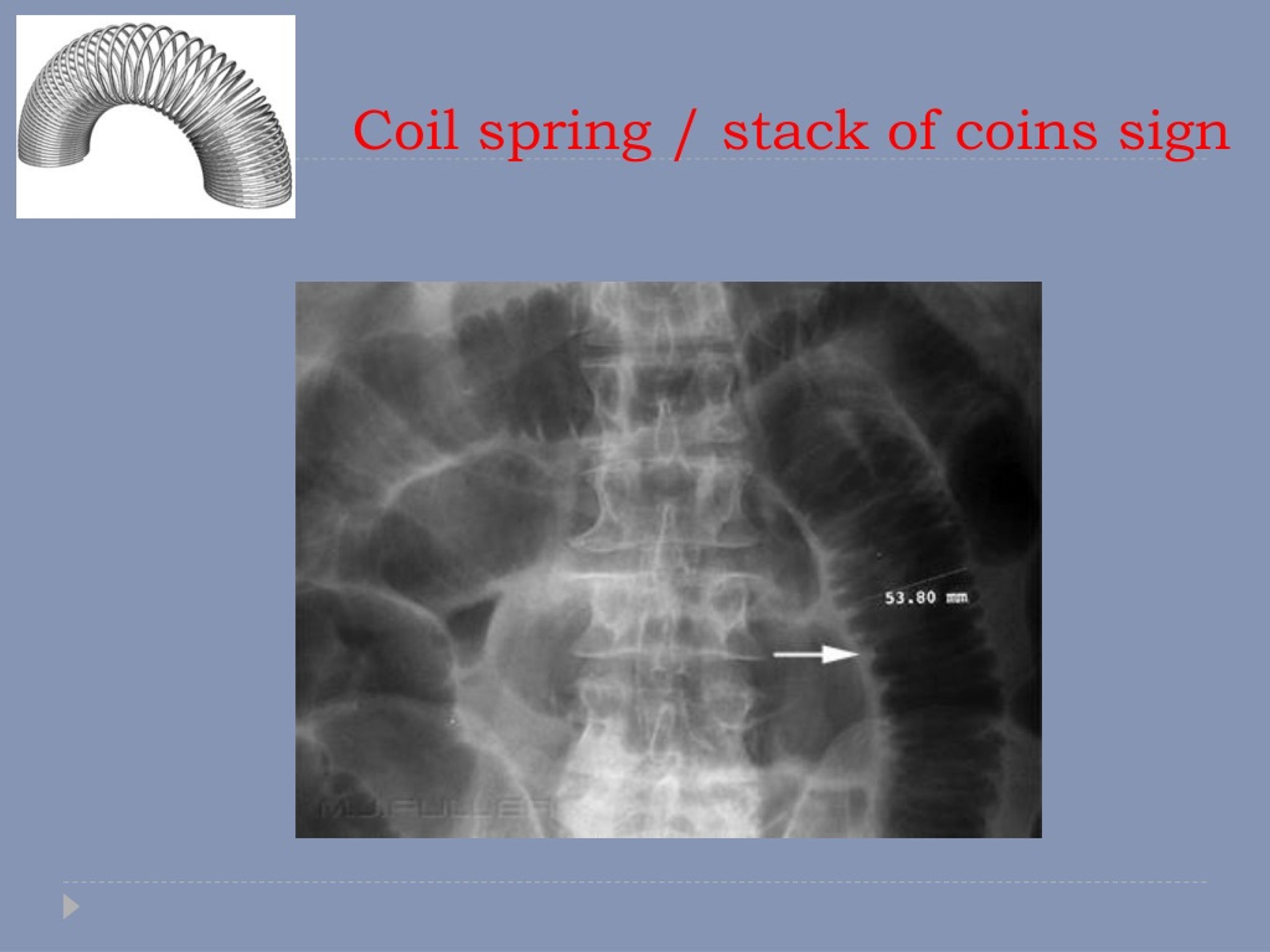
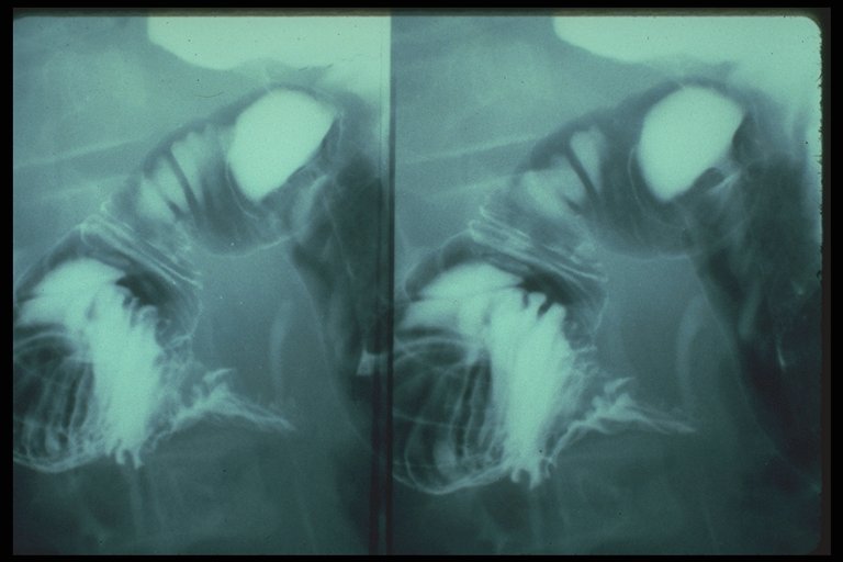
Coiled-spring sign of appendiceal intussusception Radiology. 1985 Apr;155 (1):41-4. doi: 10.1148/radiology.155.1.3975417. Authors M S Levine , S W Trenkner , H Herlinger , J D Mishkin , J C Reynolds PMID: 3975417 DOI: 10.1148/radiology.155.1.3975417 Abstract

Intussusception, Pediatric Diseases & Conditions 5MinuteConsult
Recent Advances in Radio-Frequency Coil Technologies: Flexible, Wireless, and Integrated Coil Arrays. Dean Darnell PhD,. Radio-frequency (RF) coils are to magnetic resonance imaging (MRI) scanners what eyes are to the human body. Because of their critical importance, there have been constant innovations driving the rapid development of RF.

Wholebody resonance angiography Clinical Radiology
The largest frequency shift and worst impedance matching were 3.6 MHz/−2.8 dB and 7.3 MHz/−3.2 dB for the conventional overlapped and self-decoupled coils, respectively. The normalized S21 of.
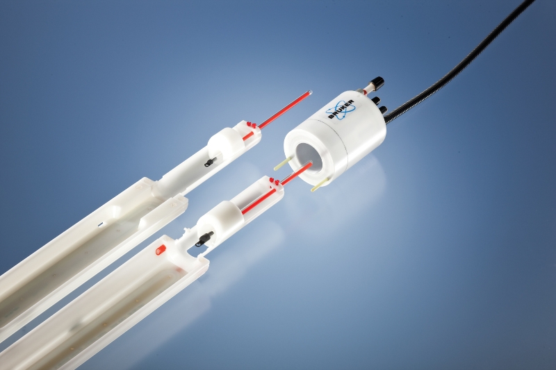
MRI RF Coil Imaging and Spectroscopy Bruker Bruker
Coil-spring embolization is a procedure for treatment of pulmonary arteriovenous malformations. Herein is described a patient with hepatogenic pulmonary angiodysplasia ("pulmonary spiders") managed with this technique. Pulmonary angiodysplasia with hepatic cirrhosis is a well-described but poorly explained entity.

Pin em DIGESTIVE
Radiofrequency (RF) coils are essential to any MRI system. They play two separate but related roles. First, specialized transmitter coils are responsible for exciting spins within the body. These coils are designed to have a uniform effect throughout the volume being imaged. A second set of RF coils are designed for receiving the MRI signal.

Simultaneous Imaging of Lung Structure and Function with TripleNuclear
The coiled spring sign refers to the double contrast enema (DCE) appearance of appendiceal intussusception into the cecum, which is best seen en face. There is non-filling of the appendix with barium, and thin concentric rings of barium radiate outward from the appendix base, (Fig. 1) giving a spring-like appearance (Fig. 2 ).
PPT Radiology of the abdomen PowerPoint Presentation, free download
Given the growing variety of specialized coils available for neuroradiologic imaging applications, it is critical that radiologists use a coherent strategy for successfully matching these coils to specific imaging situations. First, fundamental concepts of coil design are reviewed.

How DuoFLEX Flexible MRI Coils Boost MRI Scan Performance
Coiled-spring sign of appendiceal intussusception. | Radiology Home Radiology Vol. 155, No. 1 Previous Next Coiled-spring sign of appendiceal intussusception. M S Levine , S W Trenkner , H Herlinger , J D Mishkin , J C Reynolds Published Online: Apr 1 1985 https://doi.org/10.1148/radiology.155.1.3975417 PDF Tools Share Abstract
Integrated Coils Technology Resonance Imaging MRI
Various radiofrequency (RF) ablation electrode designs have been developed to increase ablation volume. Multiple heating cycles and electrode positions are often required, thereby increasing treatment time. The objective of this study was to evaluate the performance of a high-frequency monopolar induction coil designed to produce large thermal lesions (>3cm) with a single electrode insertion.
Abdominal Xray Interpretation
Radiofrequency (RF) coils are an essential MRI component used for transmission of the RF field to excite nuclear spins and for reception of the MRI signal. They play an important role in image quality in terms of signal-to-noise ratio, signal uniformity, and image resolution.
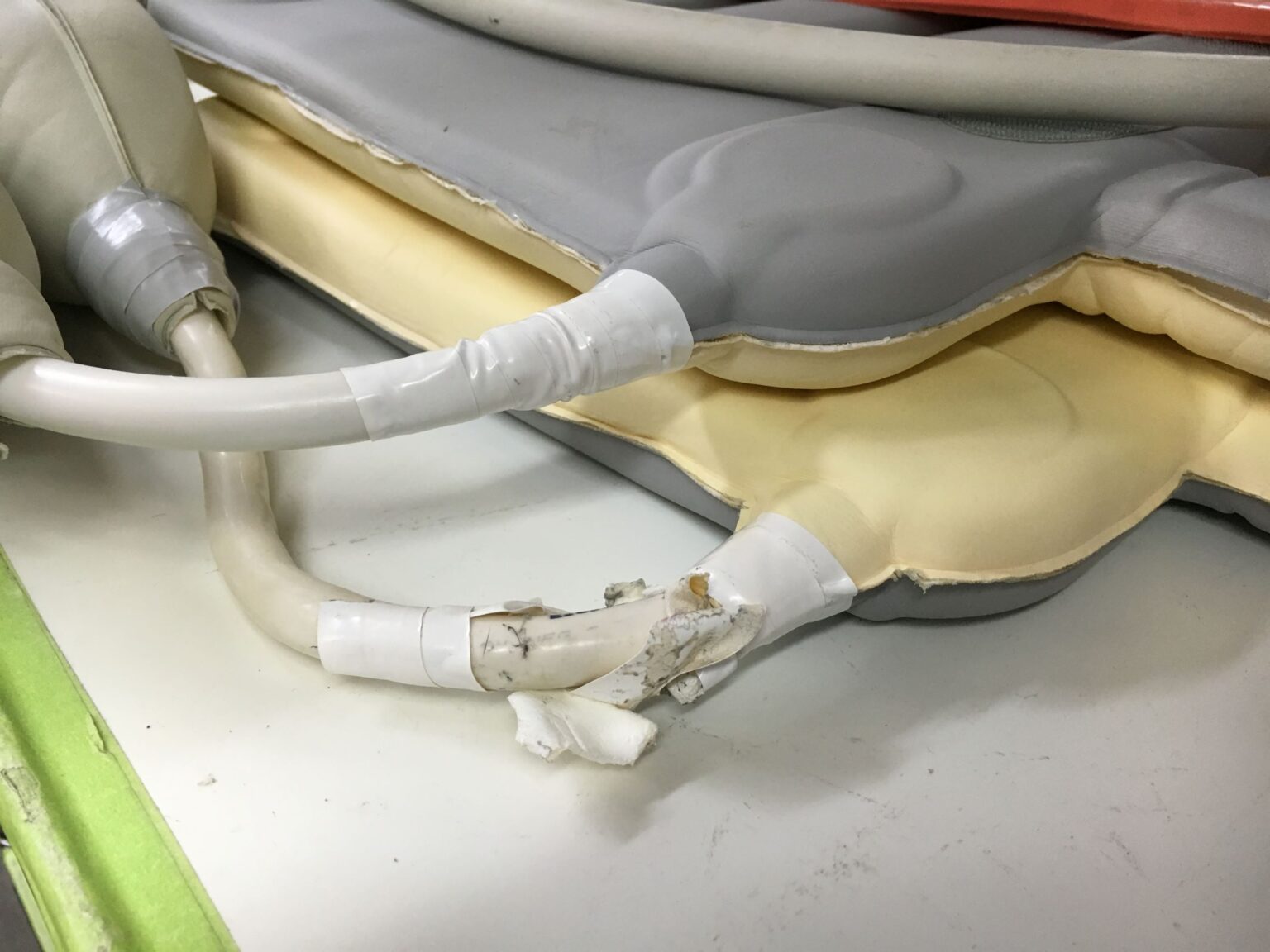
Many MRI coil failures are related to the cable Innovatus Imaging
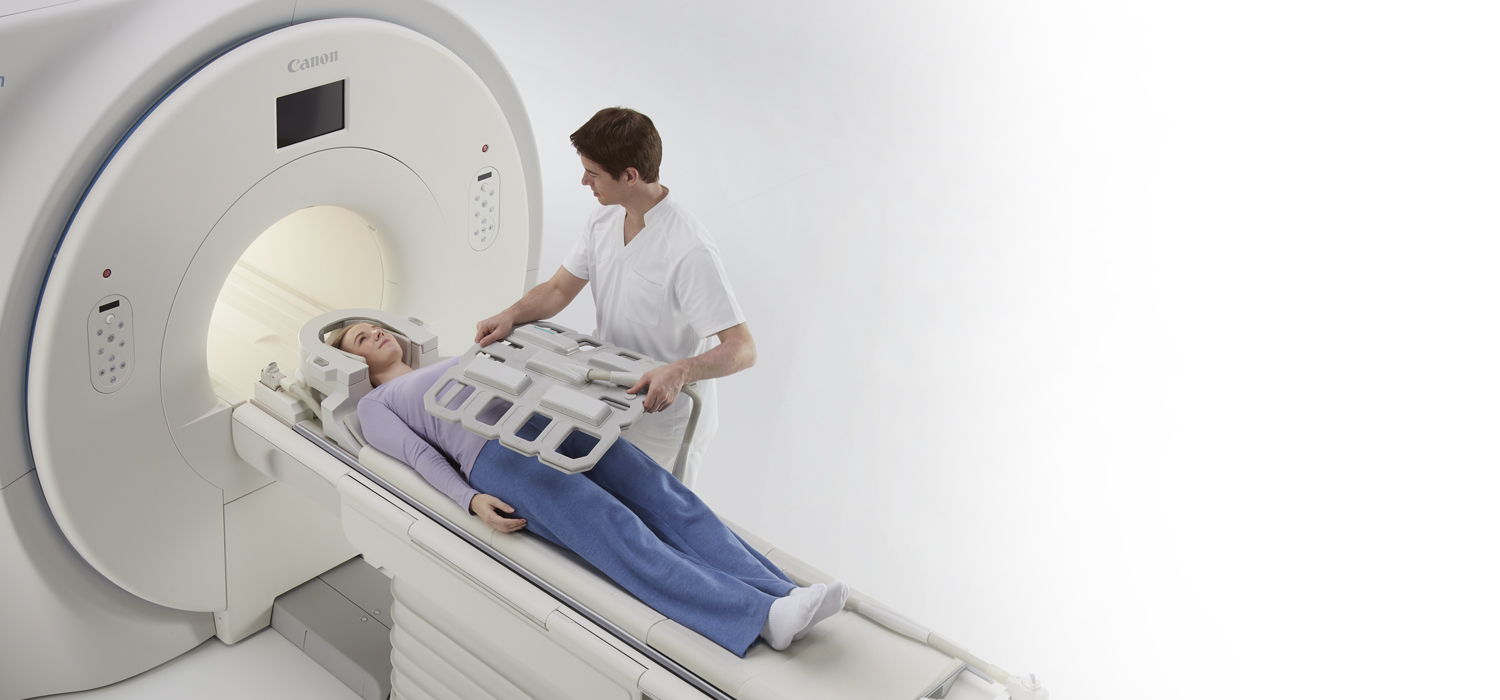
These scanners typically have spherical imaging volumes of 40-50 cm in diameter. With a subject present in the scanner, there is limited space for coil enclosures. RF coils must be in a protective housing for mechanical stability. These housings must be lightweight, non-magnetic, and non-conductive.
coiled spring sign meddic
Volume coils are the transmit and receive radiofrequency coils which are used to both transmit and receive the radiofrequency signal in MRI. Most MRI scanners have what is called a body coil - which is a volume coil built into the bore of the magnet which transmits the radiofrequency for most examinations. Certain types of imaging require.
4TIPS FOR EXTENDING THE LIFECYCLE OF MRI COILS
Radiofrequency (RF) coils are an essential MRI component used for transmission of the RF field to excite nuclear spins and for reception of the MRI signal. They play an important role in image quality in terms of signal-to-noise ratio, signal uniformity, and image resolution.

Pin on Медицинская школа
In intussusception the occluding mass prolapsing into the lumen has been termed the "coiled spring" appearance (barium in the lumen of the intussusceptum and in the intraluminal space). Related Radiopaedia articles Intussusception (advertising)

MRI Options & Upgrades Coils
The purpose of this study is to describe the sonography, CT, and MRI appearance of the Essure microinsert. Fig. 1 — Photograph of Essure microinsert (Conceptus, Inc.). Noted are two radiopaque markers at ends of inner (central) coil ( long arrows ), and two radiopaque markers at ends of outer (spring) coil ( short arrows ).

Spring ligament complex Illustrated normal anatomy and spectrum of
While the classic triad of intermittent abdominal pain, vomiting, and right upper quadrant mass, plus occult or gross blood on rectal examination, has great positive predictive value for intussusception in children 1, these findings, taken together, are seen in less than 20% of intussusception cases 2.